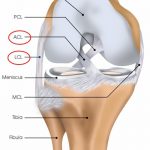
 New functional capacity evaluation for ACL injuries definitely tests likeliness for re-injury and readiness to return to sport
Home / Shoulder / Shoulder Surgery / Tendon repair / Superior Capsular Graft Augmentation of Massive Rotator Cuff
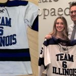
 “Thank you Dr. Chudik! I’m happy to be back on the ice.”
Dr Steven Chudik founded OTRF in 2007 to keep people active and healthy through unbiased education and research. Click to learn about OTRF’s free programs, educational opportunities and ways to participate with the nonprofit foundation.
1010 Executive Ct, Suite 250
Westmont, Illinois 60559
Phone: 630-324-0402
Fax: 630-920-2382
(New Patients)
550 W Ogden Ave
Hinsdale, IL 60521
Phone: 630-323-6116
Fax: 630-920-2382
4700 Gilbert Ave, Suite 51
Western Springs, Illinois 60558
Phone: 630-324-0402
Fax: 630-920-2382

© 2025 © 2019 Copyright Steven Chudik MD, All Rights Reserved.

